Theme: Practice, Research & Leadership: Weaving it all together
Radiology and Oncology 2019
Radiology and Oncology 2019 Congress
On behalf of the Scientific Committee, we are honored to invite you to 3rd World Congress on Oncology and Radiology to be held in the Abu Dhabi, UAE during April 8-9, 2019.
The Theme of the Radiology and Oncology -2019 is “Practice, Research & Leadership: Weaving it all together "which covers the wide range of critically important sessions in the field of Oncology, Radiology, Cancer, and Imaging. This multidisciplinary multi-specialty oncology course will cover all aspects of cancer imaging and intervention. The objective of this meeting is to share the foremost updated knowledge on the radiology and the novel therapeutic options in Cancer treatment. We will gather academicians and young inspired scientists from all around the world involved in researchers at the cutting edge in the study of the radiology and oncology.
The scientific program will consist of plenary sessions, keynote presentation, special lectures, seminars, symposiums, Oral Presentations, interactive workshop sessions, special sessions, panel discussion and Posters Presentation on the latest treatment innovations in the field of Oncology, Radiology, Medical Imaging, and Cancer by experts from both academic and business background.
Radiology and Oncology 2019 planned to achieve the knowledge transfer of highly updated and relevant information to a broad audience in oncology radiology and related specialists in the field. It can be achieved by scheduled scientific sessions, by renowned scientists and poster sessions at this radiation oncology conference, which promises to deliver something for everyone involved in cancer research or practice.
It also provides a premier interdisciplinary platform for researchers, practitioners and educators to present and discuss the most recent innovations, trends, and concerns as well as practical challenges encountered and solutions adopted in the fields of Radiology and Oncology.
Radiology and Oncology committed to creating real and reliable contributions to the scientific community. Conference Series Ltd organize 2000+ Conferences once a year across the USA, Canada, Mexico Europe, Georgia, Middle East & Asia with support from a thousand additional scientific societies and Publishes 900+ Open access journals that contain over thousands eminent personalities, putative scientists as editorial board members.
Why attend??
Encounter the target market with members from across the globe, committed to learning about Radiology and Oncology. This is the best opportunity to outreach the largest gathering of participants from around the world. Conduct presentations, distribute and update knowledge about Radiology and Oncology and receive name recognition at this 2-days event. World-eminent speakers, most recent researches, latest treatment techniques and the advanced updates in Radiation Oncology are the principal features of this conference.
Target Audience:
Our Organization would be privileged to welcome them:
- Directors of Oncology and Radiology or related Programs or Associations
- Heads, Deans and Professors of Radiology and Oncology department
- Radiology and oncology Doctors
- Business Professionals
- Radiographers and Radiology Technologists
- Educators, Scientists, and Researchers
- Research Scholar
- Oncologists
- Radiologists
- Radiation Oncologists
- Radiology Consultants
- Lab Technicians
- Healthcare professionals
- Fellows
- Residents
- Founders and Employees of the related companies
- Clinical investigators & Researcher
- Hospitals and Health Services
- Pharmaceutical companies
- Laboratory members
- Support organizers
Conference Sponsor and Exhibitor Opportunities
The Conference offers the opportunity to become a conference sponsor or exhibitor.
Highlights of latest advances in Oncology and Radiology 2019
Track 1: Oncology
Oncology is the branch of therapeutic science managing tumors, including the birthplace, advancement, determination, and treatment of harmful neoplasms. It incorporates therapeutic oncology which uses chemotherapy, hormone treatment, and diverse prescriptions to treat malignancy, radiation oncology using radiation for treatment and surgical oncology.
The oncology field has three important areas
-
Medical
-
Surgical
-
Radiation
Several types of oncology specialists usually work together to plan a patient’s overall medication and treatment plan that connects many types of procedures. For example, a patient may need medication besides a combination of surgery, chemotherapy, and radiation therapy. Here is assembled a multidisciplinary team.
-
Neuro-Oncology
-
Ocular Oncology
-
Thoracic Oncology
-
Breast Oncology
-
Gastrointestinal Oncology
-
Genitourinary Oncology
-
Gynecologic Oncology
-
Pediatric Oncology
-
Hemato Oncology
-
Molecular Oncology
-
Nuclear medicine Oncology
Track 2: Radiology Trends and Technology
Radiology is the science that uses restorative imaging to analyze diagnoses and sometimes also treat diseases inside the body. An assortment of imaging systems, for example, X-ray radiography, ultrasound, Computed tomography (CT), atomic pharmaceutical including Positron Emission Tomography (PET), and Magnetic resonance imaging (MRI) are utilized to analyze or potentially treat infections. Interventional radiology is the execution of (ordinarily negligibly intrusive) therapeutic methodology with the direction of imaging innovations.
The securing of restorative pictures is normally done by the radiographer, regularly known as a Radiologic Technologist. Contingent upon the area, the Diagnostic Radiologist, or Reporting Radiographer, at that point, translate or "peruses" the pictures and delivers a report of their discoveries and impression or conclusion.
-
Global radiology
-
Medical radiography
-
Radiation protection
-
Fluoroscopy
-
Pediatric Radiology
-
Nephrostomy
-
Spinal Cord Embolisation (AVM/DAVF)
-
Projection (plain) radiography
-
Artificial intelligence
-
Teleradiology
Track 3: Cancer Cell Biology
What is Cancer?
Cancer is caused while cells accumulate with genetic mutations anywhere in a body and begin to multiply in an uncontrolled manner. Those abnormal cells are termed cancer cells.
How does cancer begin?
Cells (building blocks of life) are the basic and smallest units of life that make up the human body. The study of cells is called cell biology. Cells multiply and grow to make new cells as the body requires them.
Normally, cells die and disappear when they become too old or faded. Then, new cells generate and take their place. Cancer occurs while genetic mutations prevent with this exact manner. Cells begin to rise uncontrollably or divide relentlessly. Certain cells may create a mass called a tumor. These abnormal cells are termed cancerous cells or tumor cells. The tumor can be cancerous or benign.
The cancerous tumor (malignant): It can grow quickly and spread to different sections of the body.
A benign tumor: This is also known as non-cancerous tumor it grow but will not spread. Several benign tumors are not dangerous to human well-being. Some kinds of carcinoma do not create a tumor. Those introduce leukemias, most kinds of lymphoma, and myeloma.
Types of cancer
Four main types of cancer are:
-
Carcinomas. A carcinoma starts in the surface of the epidermis (skin) or the tissue that coats the surface of interior organs and glands. Carcinomas ordinarily produce solid tumors. They are the most regular type of cancer. Below are some examples of carcinomas:
-
Sarcomas. A sarcoma occurs in the connective tissues that support and unite the body. A sarcoma can occur in fat, muscles, tissues, ligaments, bones or blood vessels.
-
Leukemia. Leukemia is a broader group of tumors and affected by an excess formation of damaged white blood cells and usually occur in the bone marrow. Leukemia begins when healthy blood cells change and grow uncontrollably and mutated.
The four main types of leukemia are
-
Acute lymphocytic leukemia (ALL)
-
Chronic lymphocytic leukemia (CLL)
-
Acute myeloid leukemia (AML)
-
Chronic myeloid leukemia (CML)
-
Lymphomas. A lymphoma is a group of blood cancer that starts in the lymphocytes. The lymphatic system is a network of vessels and glands that help in infection-fighting cells of the immune system. There are two main types of lymphomas:
-
Hodgkin lymphoma (HL)
-
Non-Hodgkin lymphoma (NHL)
There are many other types of cancer. Learn more about these other types of cancers.
Cancer Cell Biology
Recognition and understanding the process of cells, how cancer grows and progresses, including how gene mutations make the growth and spread of cancer cells, and how tumors communicate and interact with their enclosing circumstances and the surrounding environment, is essential for the identification of new targeted cancer treatments.
Track 4: Radiopharmaceuticals
A radiopharmaceutical is a medicine that can be used as a diagnosis and for therapeutic purposes. This is also a special class of drug and a combination of a radioactive molecule that has radioactivity. Radiopharmaceuticals consist of a radioactive isotope. Radioisotopes bind to biological molecules capable of targeting organs, tissues or specific cells of the human body. These radioactive drugs can be used for diagnosis and, increasingly, for the therapy of diseases.
The representation of radiopharmaceuticals in clinical practice is increasing rapidly, which provides the medical association to have a greater way of detailed knowledge and information on the characteristics of various kinds of tumors.
Several properties of the ideal pharmaceutical product:
• High objective: non-objective acceptance ratio
• Easy and cheap to produce
• Not Toxic
• Does not alter the physiology to give an accurate description of the patient's physiology
Track 5: Cancer biopsy
During a biopsy is the removal of a small amount of tissue in the body to examine under a microscope. It is the major way doctors diagnose most kinds of cancer. Our specialist may perform the biopsy with the help of an imaging examination. Additional tests can advise that cancer is present, but particularly a biopsy can obtain a diagnosis.
-
Image-guided biopsy
-
Fine needle aspiration biopsy
-
Core needle biopsy
-
Vacuum-assisted biopsy
-
Excisional biopsy
-
Shave biopsy
-
Punch biopsy
-
Endoscopic biopsy
-
Laparoscopic biopsy
-
Bone marrow aspiration and biopsy
-
Liquid biopsy
Track 6: Medical Imaging Technology
Medical imaging is the procedure used to make visual representations of the human body for clinical purposes or medicinal science (including the study of normal anatomy and physiology). Medical imaging technology plays an important role in the health care system.
Imaging for therapeutic purposes includes a group which includes the administration of radiologists, radiographers (X-ray technologists), sonographers (ultrasound technologists), medicinal physicists, biomedical designers, and other support staff working together to optimize the well-being of patients, each one in turn. Appropriate utilization of medical imaging requires a multidisciplinary approach.
-
Biological imaging
-
Medical ultrasonography
-
Radiography
-
Endoscopy
-
Elastography
-
Tomography
-
Photoacoustic imaging
-
Tactile imaging
-
Functional near-infrared spectroscopy
-
Single-photon emission computed tomography (SPECT)
Track 7: Pharmacokinetics
Pharmacokinetics is the study of how the body causes changes in medications (drug) and includes analysis of absorption, distribution, metabolism, and excretion. Cancer patients occupy the full spectrum of liver function, from unaffected to frank liver failure, and this invariably leads to pharmacokinetic variability
When considering a specific application of pharmacokinetics, an important consideration should be some of the other relevant aspects of the area. We will start with some general considerations of cancer chemotherapy to set the stage for the role of pharmacokinetics. This will consist of a brief description of cell kinetics.
-
Liberation
-
Absorption
-
Distribution
-
Metabolism
-
Excretion
Track 8: Radiation Oncology
Radiation oncology is one of the three basic specialties, the other two being surgical and therapeutic oncology, related to the treatment of development. Radiation can be given as a therapeutic system, either alone or in the mix with surgery or possibly chemotherapy. The mission of Advances in Radiation Oncology is to give unique clinical research went for improving the lives of people living with tumor and distinctive ailments treated with radiation treatment.
The field of radiation oncology covers the combination of radiotherapy in multimodal treatment procedures. Radiation Oncology provides an open-access study for researchers and doctors concerned in the management and treatment of cancers cases, which brings together the advanced research and analysis in the field. Advancements in processing with treatment technology, as well as a better understanding of the underlying biological defense mechanisms, will further extend the function of radiation oncology. A radiation oncologist is a medical specialist who uses radiation therapy in the treatment of patients with cancer.
Specialists who use this technology or the knowledge concerned from it involve:
-
Radiation oncologists
-
Radiation therapists
-
Radiation oncology nurses
-
Medical radiation physicists
-
Dosimetrists
-
Social workers
-
Dietitians
-
External beam radiation therapy
-
Proton therapy
-
Brachytherapy
-
Chemotherapy
-
Stereotactic radiosurgery
-
Radioembolization
Track 9: Anesthesia
Anesthesia is a method to control pain during a surgery or procedure by using the medicine called anesthetics. It can help control your breathing, blood pressure, blood flow, and heart rate and rhythm.
Anesthesia may be used to:
-
Relax you
-
Block pain
-
Make you sleepy or forgetful.
-
Make you unconscious for your surgery
Track 10: Oncology Nursing and Care
Nursing is a profession within the health division concentrated on the care of people, and societies so that they can gain, have or obtain optimal well-being and quality of life. Nurses generate a care program, working in collaboration with doctors, therapists, the patient, the patient's family, and other organization members, which concentrates on treating the disease to enhance the quality of life.
An oncology nurse is a specialized nurse who treats cancer patients. Oncology nursing care can be defined as satisfying the diverse needs of oncology patients during the time of their illness, including adequate screening and other preventive practices, symptom management, care to maintain the highest possible normal functioning and measures of support at the end of life.
Oncology nurses can also work in inpatient environments, such as hospitals, clinics, outpatient clinics, or doctor's offices. There are a variety of specialties such as radiation, healthcare, surgery, pediatrics or gynecology. The oncology nurses have superior information and awareness of the evaluation of the client's situation and this evaluation will support the multidisciplinary medical team to occur a treatment program.
-
Chemotherapy and biotherapy
-
Nursing Informatics
Track 11: Cancer Awareness
Cancer is the uncontrolled growth of abnormal cells in the body. Cancer is one of the leading causes of death worldwide. It represents 8.5 million deaths (around 15% of all deaths) in 2017. The majority of cancers, around 80-85% of cases, are due to genetic mutations caused by environmental factors. The remaining 10-15% is due to inherited genetics.
Environmental, as practiced by tumor researchers, determines some reason that is not genetically inherited, such as lifestyle, commercial and behavioral circumstances and factors, and not only pollution.
Common environmental factors that contribute to death from cancer include tobacco (30-40%), diet and obesity (20-30%), infections (10-20%), radiation (both ionizing and non-ionizing, up to 15%), stress, lack of physical activity and pollution.
Cancer usually generates fear that originates from ignorance and error. More than 40% of cancer patients could be prevented by changing lifestyle or by avoiding key risk agents. Approximately 1/4 of cancer cases could be reduced if cases are treated and detected at an early stage.
Objectives of cancer awareness
To create awareness of the disease.
Help people recognize the early signs and symptoms of cancer, allowing them to seek treatment at an early stage. The presentation support and encourages participants to explore prompt medical care for signs that may involve lumps, wounds, bleeding, hoarseness, weight loss, and persistent indigestion/ a cough/pain, etc.
Educate people about the main risk factors for cancer, since more than 40% of cancer cases could be prevented by modifying lifestyle or avoiding key risk factors.
Inform people about the importance of cancer screening at an early stage and motivate them to take over cancer control services at a very nominal cost through the Cancer Detection Center.
Achievements of cancer awareness
During 2010-2017, 190 educational and awareness-raising programs on cancer were carried out in various places, such as schools, institutes, private companies, NGOs and government organizations. A total of 30,315 participants benefited from these programs. During the programs, information was provided on cancer, the causes, and symptoms of cancer, types of cancer, cancer control, and its importance, treatment, and prevention of cancer. During the program, pamphlets related to cancer were distributed among the participants to educate them about cancer.
Track 12: Nuclear Medicine
Nuclear Medicine is a medical specialty that utilizations radioactive tracers (radiopharmaceuticals) to distinguish substantial capacities and to analyze and treat an infection. Nuclear medicine is a branch of radiology and therapeutic imaging. The unique way to kill malignancy cells with insignificant damage to encompassing tissue.
Nuclear medicine treatment utilizes a larger amount of radiation to treat thyroid disease and tumor. It utilizes a smaller number of radioactive pharmaceuticals. This method help characterizes diseases in practically every organ system including the heart, mind, skeleton, thyroid and kidneys and many types of tumor and can be utilized to treat illness without surgery. This is one of a unique approach to kill cancer cells with minimal damage to surrounding tissue.
Track 13: Interventional Radiology
Interventional radiology also is known as vascular and interventional radiology (VIR), is a medical specialty which provides minimally invasive image-guided diagnosis and treatment of disease. Although the procedure range performed by interventional radiologists is broad, the unifying concept behind these procedures is the application of image guidance and minimally invasive techniques in order to minimize risk to the patient.
Interventional radiologists perform a wide range of procedures, including:
-
Image Guided Cervical Nerve Root Sleeve Corticosteroid Injection
-
Carpal Tunnel Ultrasound and Injection
-
Image-Guided Liver Biopsy
-
Bursal Injection
-
Image-guided lumbar nerve root sleeve injection
-
Biliary Drainage
-
Angioplasty and Stent Insertion
-
Ascitic Tap
-
Uterine Fibroid Embolisation
Track 14: Cancer pharmacology
Cancer pharmacology is a peer-reviewed medical journal covering oncological pharmacotherapy and it involves studies of the basic mechanisms of signal transduction associated with cell proliferation and apoptosis (cell program death), the mechanisms of action of antineoplastic agents, the design and development of new drugs, basic mechanisms of DNA repair and tolerance to cell damage. DNA and the development of strategies for gene therapy
Importance is arranged on the classification and characterization of the basic cell signaling mechanisms that create the targets of the molecules worked for cancer therapy and DNA destruction and repair mechanisms that provide resistance to antineoplastic drugs.
The regulation of tyrosine kinases, the processing of proto-oncogenes, the regulation of small GTPases and their effectors, the specific kinases of the cell cycle and the products of the DNA repair gene are being studied as potential targets or to improve the efficacy of chemotherapeutic agents existing The role of growth factors in the progression of solid and hematopoietic tumors is being studied; New receptors and signal transduction pathways are being identified in normal and malignant tissues.
Track 15: Breast Cancer-Present Perspective
At least one in nine women develops breast cancer at some stage in their life. About 48,000 cases occur in the United Kingdom every year. Mostly develops in women over the age of 50 but younger women are also sometimes affected. Breast cancer can also develop in men, but this is rare. Breast cancer develops from a cancerous cell which develops in the lining of a mammary duct or a lobule in one of the breasts. It follows the classic progression though it often becomes systemic or widespread in the early onset of the disease. During this period, cancer may metastasize, or spread through lymphatics or bloodstream to areas elsewhere in the body. If breast cancer spreads to vital organs of the body, its presence will compromise the function of those organs. Fetal death is the result of an extreme case of vital organ function. In 2012, the latest year for which information is accessible, in excess of 1.7 million ladies worldwide were determined to have Breast Cancer.
A large number of these findings are influenced utilizing X-to beam mammography. Albeit standard and broadly utilized, X-ray imaging for breast cancer experiences both low affectability (50-75%) and the utilization of ionizing radiation that can't be thought about totally safe at least one in nine women develops breast cancer at some stage in their life.
About 48,000 cases occur in the United Kingdom every year. Mostly develops in women over the age of 50 but younger women are also sometimes affected. Breast cancer can also develop in men, but this is rare. Breast cancer develops from a cancerous cell which develops in the lining of a mammary duct or a lobule in one of the breasts. It follows the classic progression though it often becomes systemic or widespread in the early onset of the disease.
During this period, cancer may metastasize, or spread through lymphatics or bloodstream to areas elsewhere in the body. If breast cancer spreads to vital organs of the body, its presence will compromise the function of those organs. Fetal death is the result of an extreme case of vital organ function. In 2012, the latest year for which information is accessible, in excess of 1.7 million ladies worldwide were determined to have Breast Cancer.
A large number of these findings are influenced utilizing X-to beam mammography. Albeit standard and broadly utilized, X-ray imaging for breast cancer experiences both low affectability (50-75%) and the utilization of ionizing radiation that can't be thought about totally safe.
Track 16: Ultrasound
Ultrasound is a sort of imaging and part of radiology. It utilizes high-frequency sound waves to catch live images from inside your body. Ultrasound is protected and easy. Ultrasound is a valuable methodology for observing the child's improvement in the uterus.
-
Abdominal Ultrasound Imaging
-
Pelvic Ultrasound Imaging
-
Obstetric Ultrasound Imaging
-
Ultrasound attenuation spectroscopy
-
Medical ultrasonography
Track 17: Cancer Prevention and Research
Cancer study and research to recognize causes and develop advanced strategies for prevention, inhibition, diagnosis, medication, treatments, and cure. These applications involve surgery, radiotherapy, chemotherapy, hormonal therapy, immunotherapy and merged therapy and treatments modalities such as chemo-radiotherapy and radiotherapy.
Cancer prevention is an action that is taken to reduce the risk of getting cancer. This can include maintaining a healthy lifestyle, avoiding exposure to known substances that cause cancer, and taking medications or vaccines that can prevent the development of cancer.
Here are some highlights for cancer prevention
- Be as thin as possible without losing weight.
- Stay physically active for at least 40 minutes every day.
- Avoid sugary drinks and limit the consumption of high-calorie foods, especially those that are low in fiber and high in added fat or sugar.
- Have more than one category or a variety of vegetables, fruits, whole cereals, grains, and legumes.
- Limit the consumption of red meats involving beef, pork, and lamb and avoid prepared meats.
- Limit your everyday intake to two drinks for men and one drink for women, while you drink alcohol.
- Limit the consumption of salty foods and prepared meals with salt.
- Do not use additions supplements to try to defend against cancer.
- Later treatment, carcinoma survivors should obey the instruction and recommendations for cancer prevention
- The best thing for mothers is to exclusively breastfeed their babies for up to six months and then add other fluids and foods.
Track 18: Positron Emission Tomography/Computed Tomography- PET/CT/X-ray
Positron-outflow tomography (PET) is an atomic medication utilitarian imaging strategy that is utilized to watch metabolic procedures in the body. The framework recognizes sets of gamma beams discharged in a roundabout way by a positron-transmitting radionuclide (tracer), which is brought into the body on a naturally dynamic atom. In present-day PET-CT scanners, three-dimensional imaging is frequently refined with the guide of a CT X-beam filter performed on the patient amid a similar session, in a similar machine.
Computed tomography (CT), sometimes called "computerized tomography" or "computed axial tomography" (CAT), is a noninvasive medical examination or procedure that uses specialized X-ray equipment to produce cross-sectional images of the body.
- CT is a valuable medical tool that can help a physician:
- Diagnose disease, trauma or abnormality
- Plan and guide interventional or therapeutic procedures
- Cancer treatment
Track 19: Magnetic Resonance Imaging (MRI)
Magnetic resonance imaging (MRI) of the body uses a powerful magnetic field, radio waves and a computer to produce detailed pictures of the inside of your body. It may be used to help diagnose or monitor treatment for a variety of conditions within the chest, abdomen, and pelvis.
-
Magnetic resonance angiography (MRA)
-
Cardiac MRI
-
Magnetic resonance venography (MRV)
-
Breast scans
Track 20: Nanotechnology in Cancer Treatment
Nanotechnology is rapidly developing a technology subdivision that affects many fields. Nanoscale devices are one hundred to ten thousand times smaller than human cells. They are similar in size to large biological molecules, such as enzymes and receptors. Due to their small size, nanoscale devices can easily interact with biomolecules both on the surface and inside cells.
Medicine is also affected by nanotechnology; since, in the treatment of cancer, nanotechnologically modified methods can be used. One of the fields of use in the development of nanotechnology is the treatment of cancer.
Nanotechnology can help to have a better diagnosis with less harmful substances such as optical nanoparticles. A subdivision of technology that is nanotechnology will play an important role. Nanotechnology can provide rapid and sensitive detection of cancer cells, a more effective administration of drugs to tumor cells, a molecularly targeted cancer therapy, and highly effective therapeutic agents.
Track 21: Cancer Therapies
Cancer Treatments are medical therapies that asserted to treat cancer through different methods like surgery, chemotherapy, radiation oncology, and immunotherapy. Oncolytic biotherapy is a rising treatment method of cancer which uses infections to destroy cancers. The recent development in genetic engineering techniques has been made using viruses to attack and destroy cancer cells. Chemotherapy is a method of cancer treatment which uses chemical substances or chemotherapeutics drug to kill the cancerous cells. Radiologists play an important role not only in developing and mastering endovascular genetic invasions but also in evaluating the progress of vascular gene therapy and reading further cultivation of vascular gene therapy technology.
It is one of the major methods of medical oncology.
Several types of radiation treatment are used to treat cancer.
-
Radiotherapy and Chemotherapy
-
Immunotherapy
-
Stem cell Transplantation
-
Hormone Therapies
-
Genomic Tumor Assessment
-
Precision Medicine
-
Chemotherapy
-
Stereotactic Radiosurgery
-
Molecular Targeted Cancer Therapies
-
HDR Brachytherapy
-
External Beam Radiation
Track 22: Neuroradiology and Neuro-oncology
Neuroradiology is the subdivision of radiology that deals with the sensory or nervous system. It is the subspecialty of radiology which is converging on the diagnosis and characterization of variations from the peripheral nervous system, spine, and head and neck utilizing neuroimaging strategies. X-rays are utilized as a part of the analysis and treatment of nervous system disorders. Essential imaging modalities incorporate Computed tomography (CT) and Magnetic resonance imaging (MRI). Ultrasound and Plain radiography are used on a constrained premise and in limited conditions, individual in the pediatric population.
Neuroradiology has a critical part to play in the Diagnosis and Treatment of Several Neurological issues like Ischemic Stroke and the Structural injuries causing Cerebral Haemorrhage. The current advances are rising all the more quickly in the exploration fields of Neuroradiology incorporates the improvement of MR imaging of the Brain and Spinal cord neoplasms.
-
Brain Tumor
-
Clinical Neuroscience
-
Neurosonology
-
Radiation Technology in Neuroscience
-
Brain Morphometry
-
Clinical Neuroradiology
-
Spine Intervention
-
Neuroinflammation
-
Central Nervous System Malignancies
-
Whole-brain radiotherapy (WBRT)
-
Multivariate Deposits In Brain Imaging
Track 23: Biomarkers and Cancer targets
Biomarkers or the molecular markers are the biological molecules (such as DNA, RNA, and Proteins), genes or the processes such as apoptosis and proliferation by which a specific disease, condition or any abnormalities in the body can be identified. It is a measurable indicator in a biological system and can be found in blood, tissues, specific organs, cell lines and in other body fluids.
They play a major role in the diagnosis as well as for the management of Cancer. Cancer biomarkers are either produced by the tumors itself or in response to the disease or other associated factors such as inflammation, indicating the presence of the Cancer in the body.
Biomarkers are primarily used in three ways for Cancer research and medicine:
- Diagnostic (or screening) biomarker: For the diagnosis of the condition, as in the case of an early stage of the Cancer.
- Prognostic biomarker: forecasting of the condition that how aggressive it is and determining the patient’s response in the absence of the treatment.
- Stratification (predictive) biomarker: to predict the patient’s response to the treatment.
When is radiation therapy used?
Radiation treatment devising is meant to ensure that the radiation therapy will have supreme benefits with minimum inherent risk. This involves working out an exact site, an angle of radiation, dose, and so on.
Radiology and Oncology 2019 estimates that around 70% of cancer patients receive radiation therapy as part of their treatment at some point.
Cancers that can be particularly suitable for radiation therapy aimed at curing the disease are those that are well-defined and confined. This allows the complete range of malignant tissue to be targeted by the radiation. In contrast, some modes of cancer - lymphoma or leukemia, say - can be treated with total body irradiation. Radiation therapy can be used to decrease indications, begun by tumor growth. Radiation contributes an option to the replacement of the vocal chords, avoiding surgical wound while holding a similar efficiency as surgery.
Radiation therapy can be used adjacent to both surgery and chemotherapy. Sarcomas or tumors of the breast, esophagus, lung or rectum may be treated with all three modalities.
Radiation planning can be a complete process comprising of a number of health-care experts, including researchers and consultants (radiologists and oncologists), nurses, radiographers and other technicians at the 3rd world congress on Radiology and Oncology going to be held at Abu Dhabi, UAE during April 08-09, 2019.
For more visit https://radiology-oncology.annualcongress.com/
Handheld probe images photoreceptors in children
Researchers have developed a hand-held probe that can obtain images of individual photoreceptors in babies' eyes. The technology, based on adaptive optics, will make it easier for doctors and researchers to observe these cells to diagnose eye diseases and make early detection of diseases and traumas related to the brain. Photoreceptors are specialized neurons that comprise cells that are sensitive to the light of the retina, an extension of the central nervous system located in the back of the eye. The retina sends signals to the brain through the optic nerve, which then processes the visual information. Earlier examinations have revealed that neurodegenerative disorders, like as Alzheimer's, as well as traumatic brain injuries and disorders, such as concussions, can treat the neural structures of the retina.
After studies, professionals commonly work with AOSLO (adaptive optical scanning laser ophthalmoscope), that is a non-invasive device that provides the higher resolution as the comparison of magnetic resonance imaging (MRI).
Other researchers are saying that the wavefront sensor can be replaced by an algorithm because previous algorithms have not been fast and active or robust enough to be used in a handheld method.
The algorithm we develop is much faster than the previous techniques and just as accurate. The tool was tested in a clinical trial with some adults and children, where the team demonstrated its ability to capture detailed images of photoreceptors near the fovea: the center of the retina where the photoreceptors are smaller and the vision sharper.
Our new tool is fast and light so that doctors can take it directly to their patients, and the probe allows us to collect images quickly, even if there is movement. These capabilities allow us to open the group of patients who could benefit from this technology. Before researchers prepare for large-scale clinical trials, they plan to incorporate additional imaging modes to detect other diseases
Scientists have identified a new enzymatic mechanism that induces cancer cells that are about to migrate to destroy themselves by degrading their small power plants or mitochondria. They hope that this discovery will lead to new treatments that can stop the spread of tumors.
How key cancer-related enzyme functions
Researchers have observed how an enzyme works that plays a key role in the development of cancer. The researchers hope that the new knowledge will lead to the design of more precise medications. The new study shows, first, that the METTL13 enzyme helps control the formation of new proteins in cells. Errors in protein production are undesirable, as we know from other studies that it can result in the development of cancerous tumors and degenerative brain disorders.
The enzyme helps control protein synthesis by placing the so-called methyl labels on a particular protein called eEF1A. If methyl is not properly connected to the protein, it cannot do its function properly. And that also affects the formation of proteins in the cell, which will take place at a suboptimal rate, and although that does not sound so bad, it seems to be related to very serious disorders.
Researchers have shown how the enzyme works by studying isolated human cancer cells using advanced mass spectrometers that, in short, are able to identify and quantify proteins and their methyl modifications in a cell.
Enzymes have two functions:
1. Stop the division and spread of cancer cells
2. The cancer cells are coated with 17 layers of fibrin that prevent the immune system from detecting them. High doses of enzymes will "go through" that protective layer and make cancer cells more vulnerable.
Now the researchers hope that the study will lead to the development of methods and drugs capable of targeting and ensuring that the enzyme works as expected in our cells.
Mitochondrial linkage of Cancer
Recently, from the studies of aggressive breast cancer cells, scientists have found that the movement of mitochondria inside the cancer cell is being controlled by the pathway- called Arf6-AMP1-PRKD2. It is known that the mitochondria generally move inside the cells under various circumstances but in cancer cells, it congregates in the leading rim of the cell. It is due to the pathway- Arf6-AMP1-PRKD2, which helps to recycle the protein, named integrin in the cancerous cells which then forms the adhesion complex in the cell’s membrane triggering the movement of mitochondria to the edge of the cell.
To understand the motion of the mitochondria in the cells, the researchers carried out the experiments in the breast cancer cells where they blocked the pathway. They observed that instead of the edge of the cell, the mitochondria congregate in the middle of the cell and thus, reduced invasiveness. This happens because the mitochondria started to produce and release excessive amounts of unstable oxygen-rich molecules known as reactive oxygen species (ROS) in the middle of the cell. The ROS molecules promote cancer invasiveness up to certain levels but when they become in excess, they kill cancer cells.
Radiotherapy uses ionizing radiation to shrink or eradicate tumors by increasing the production of ROS molecule inside the cancer cells. However, some cancers have become resistant to radiotherapy and other treatments that employ the ROS molecules as they have developed the tolerance to the molecules. But these findings of the molecular link between cell movements and mitochondrial dynamics may lead to novel strategies to improve ROS-mediated cancer therapies.
Happy you, Healthy you.
Chronic stress boosts cancer cell growth. Recent studies have proved that the chronic stress cause elevation in hormone epinephrine which acts directly on the cancer stem cells progression.
The Market Analysis Report on Radiology and Oncology is expected to reach USD 25.50 Billion by 2020 from USD 17.99 Billion in 2017. The market is broadly classified into the product, procedure type, and application.
The product segment of the market is further divided into Radiography system, angiography systems, fluoroscopy systems, CT scanners, ultrasound imaging systems, MRI systems, and other medical devices.
Radiology and Oncology markets continue to grow amid a more educated global population, increased awareness and advanced technology used for Disease diagnostics.
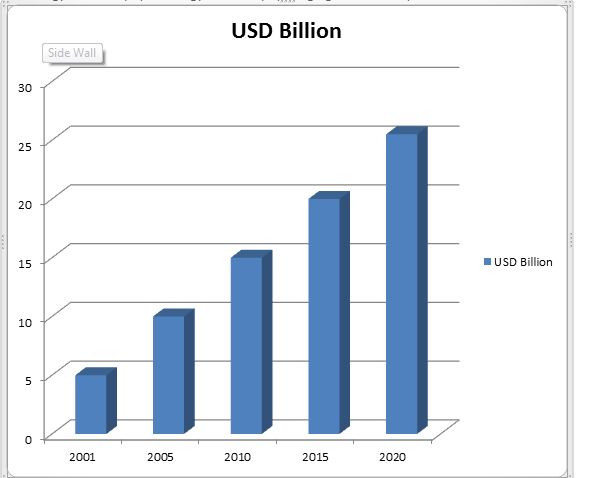
Market Analysis Report on Radiology and Oncology in USD Billion
The number of new cancer cases diagnosed every year is increased from 14.9 million in 2012 to 25.50 million by 2020. The expansion in new cases is because of a steadily aging population. Both developed and developing countries have a maturing or growing population.
Innovation and technology enhance the accuracy and appropriateness of radiotherapy and radiosurgery. Large number uses of Radiotherapy and radiosurgery equipment’s occur because the units are able to treat a broader range of cases. Advances in equipment and programming are providing a business opportunity replacing an aging installed base. New designs and outlines can convey high standards of care.
The rise in cancer cases, together with the expansion in the sophistication of new treatment etiquette, has made interest in more automated products. Automation relies upon incorporation of a few devices into clinically useful frameworks. Coordinated frameworks make medications quick and practical.
Innovation progresses prompt enhancements in patient care. The accessibility of advanced, mechanized and productive clinical apparatuses in radiation therapy has brought more exact types of radiotherapy treatment (IMRT, IGRT, VMAT, SRS, SBRT, brachytherapy and proton treatment). Innovation and Technology incorporate the EDGE™ and Truebeam™, and the Accuray TomoTherapy H Series and CyberKnife M6 stages that empower medications that diminish treatment times and increment persistent throughput.
Target Audience for this Report:
- Radiology and Oncology manufacturer and distributors
- Medical Institutions (Hospitals, Healthcare, Medical centers, Diagnostic centers Medical schools, Group practices, Individual surgeons, and Research labs) various research centers and consulting companies
Global Interventional Radiology Market: Overview
The report on the global interventional radiology market examines the current and future growth prospects of that market globally. The aim of the research statement is to give a satisfying analysis of the growth trends and the competitive structure of the interventional radiology market for the 2017-2025 estimate period. Stakeholders in the report include companies and intermediaries involved in the manufacture of imaging equipment such as x-rays and ultrasound. The section also provides comprehensive information on the global interventional radiology market based on product, application, end user, and geography. at a regional and global level.
Market revenues have been provided in terms of value (US $ Mn) and volume (in kilotons) for the global interventional radiology market for the forecast period 2018-2025. Income forecasts have also been provided for all segments and sub-segments with 2017 as the base year.
Related Conferences
- International Conference on Biomarkers and Cancer Targets October 14-15, 2019 Abu Dhabi, UAE
- 9th World Conference on Women’s Health and Breast Cancer December 09-10, 2019 Dubai, UAE
- International Conference on Metabolic Diseases and Liver Cancer, May 27-28, 2019 Istanbul, Turkey
- 26th Cancer Genomics Congress: New Era for Cancer Prevention, July 15-16, 2019 Abu Dhabi, UAE
- International Conference on Biomarkers and Cancer Targets, October 14-15, 2019 Abu Dhabi, UAE
- 21st World Congress on Radiology and Oncology May 30-31, 2019 Orlando, Florida, USA
- 3rd International Conference on Cancer Biology, Anti-Cancer Therapies and Drug Development & Delivery May 30-31, 2019 Orlando, Florida, USA
- 16th Asia Pacific Oncologists Annual Meeting May 13-14, 2019 Holiday Inn Singapore Atrium, Singapore
- 3rd World Congress on Advanced Cancer Science & Therapy August 26-27, 2019 Osaka, Japan
- 2nd Global Meeting on Clinical Oncology and Radiology March 27-28, 2019 Hong Kong
- 7th World Congress and Expo on Oncology & Radiology December 06 - 07, 2019 Kuala Lumpur, Malaysia.
- 31st Experts Meet On Cancer Therapy September 13-14, 2019 Holiday Inn Singapore Atrium, Singapore
- Society of Radiologists in Ultrasound Annual Meeting October 3 - 6, 2019. Washington DC, USA
- 6th World Summit on Cancer Research & Therapy August19-20, 2019, Dubai, UAE.
- 16th International Congress on Radiation Research August 25 - 29 2019 Manchester, UK
- 34th International Conference on Oncology Nursing and Cancer Care, July 29-30, 2019 Melbourne, Australia
Related Societies
Middle East
- Emirates Radiology Society
- Pan Arab Interventional Radiology Society
- Turkish Society of Radiology
- Middle East Cancer Consortium
- Arab Medical Association Against Cancer
USA
- Society of Nuclear Medicine and Molecular Imaging
- American Board of Radiology
- Association of Educators in Imaging and Radiologic Sciences,
- The Association for Medical Imaging Management
- American Institute of Ultrasound in Medicine
- American Society of Head and Neck Radiology
- American Society of Neuroradiology
- Canadian Association of Radiologists
- American College of Nuclear Medicine
- American Society of Clinical Oncology
- American Association for Cancer Research
- Society for Neuro-Oncology
- Mexican Association of Ultrasound in Medicine
- American Osteopathic College of Radiology
- American Society for Radiation Oncology
- American Cancer Society, American Society of Pediatric Haematology/Oncology
- Association of Community Cancer Centers, Musculoskeletal Tumour Society
- National Cancer Registrars Association
- Oncology Nursing Society
- Society for Immunotherapy of Cancer
- Long Island Radiological Society
Europe
- Italian Society of Medical Radiology
- European Society for Medical Oncology
- Argentinian Federation of Diagnostic Radiology and Radiation Therapy Associations
- European Society for Therapeutic Radiology and Oncology
- European Society of Head and Neck Radiology
- European Society of Cardiac Radiology
- Eastern Radiological Society
- Computerized Medical Imaging Society
- Cardiovascular and Interventional Radiological Society of Europe
- European Society of Musculoskeletal Radiology
- French Society of Radiology
- French Society of Nuclear Medicine
- Spanish Society of Nuclear Medicine
- Spanish Society of Vascular and Interventional Radiology
- Spanish Society of Radiology
- Nordic Society of Medical Radiology
- Norwegian Society of Nuclear Medicine and Molecular Imaging
- Swedish Society of Nuclear Medicine
- Swiss Society of Nuclear Medicine
- Swedish Society of Radiology
- British Nuclear Medicine Society
- Society of Radiologists in Training
- British Institute of Radiology
- British Society of Neuroradiologists
- British Society of Interventional Radiology
- Long Island Radiological Society
- Swiss Congress of Radiology
- Czech Radiological Society
- Czech Society of Nuclear Medicine
- Czech Society for Ultrasound in Obstetrics and Gynaecology
- Society of Diagnostic Medical Sonography
- Society for Vascular Ultrasound
- European Society of Head and Neck Radiology
Asia-Pacific
- Hong Kong Anti-Cancer Society
- Society of Indian Radiographers
- Indian Cancer Society
- Korean Cancer Association
- Asian Clinical Oncology Society
- Japan Radiological Society
- Japan Society of Clinical Oncology
- Japan Radiological Society
- Japan Medical Imaging and Radiological Systems Industries Association
- Japanese Society for Magnetic Resonance in Medicine
- Japanese Society of Sonographers
- Japanese Society for Radiation Oncology
- Japanese Society for Therapeutic Radiology and Oncology
- Japan Society for Molecular Imaging
- Society of Nuclear Medicine India
- Chinese Society of Nuclear Medicine
- International Society for Therapeutic Ultrasound
- Radiological Society of the Republic of China
- Asia Pacific Society of Cardiovascular & Interventional Radiology
- Hong Kong Society of Nuclear Medicine
- Korean Society of Radiology
Conference Highlights
- Oncology
- Radiology Trends and Technology
- Cancer Cell Biology
- Cancer biopsy
- Medical Imaging Technology
- Pharmacokinetics
- Radiation Oncology
- Anaesthesia
- Oncology Nursing and Care
- Cancer Awareness
- Nuclear Medicine
- Interventional Radiology
- Cancer pharmacology
- Breast Cancer-Present Perspective
- Ultrasound
- Cancer Prevention & Research
- Positron Emission Tomography/Computed Tomography- PET/CT/X-ray
- Magnetic Resonance Imaging (MRI)
- Nanotechnology in cancer Treatments
- Radiopharmaceuticals
- Neuroradiology and Neuro-oncology
- Cancer Therapies
- Clinical Radiology
- Biomarkers and Cancer Targets
- Radiation Protection
- Artificial Intelligence in Radiology
- Hematology-Oncology
To share your views and research, please click here to register for the Conference.
To Collaborate Scientific Professionals around the World
| Conference Date | April 08-09, 2019 | ||
| Sponsors & Exhibitors |
|
||
| Speaker Opportunity Closed | Day 1 | Day 2 | |
| Poster Opportunity Closed | Click Here to View | ||
Useful Links
Special Issues
All accepted abstracts will be published in respective Our International Journals.
- Journal of Radiology
- International Journal of Clinical & Medical Imaging
- Journal of Cancer Science & Therapy
Abstracts will be provided with Digital Object Identifier by


